What is the difference between gamma camera and PET scan?
Gamma cameras and PET (positron emission tomography) scans are both types of medical imaging that use radionuclide tracers to produce images of the body’s organs and tissues. These imaging modalities are commonly used in the diagnosis and management of a variety of medical conditions, including cancer, cardiovascular disease, and neurological disorders. While both techniques involve the injection of a radionuclide tracer into the patient’s body, they differ in the type of detector used to detect the emitted radiation and the level of detail in the resulting images. In this article, we will explore the differences between gamma cameras and PET scans in more detail.
What is a gamma camera?
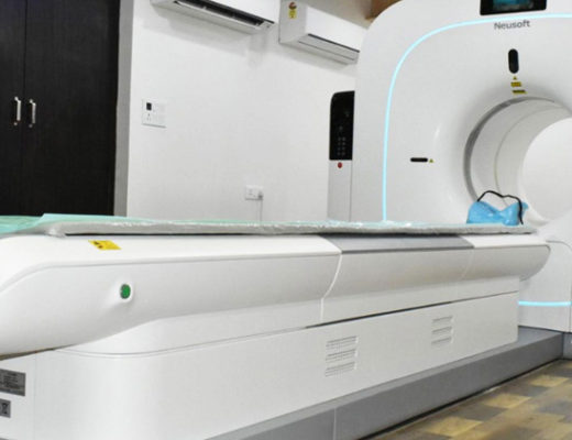
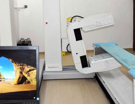
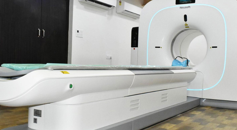
A gamma camera, also known as a scintillation camera, is a type of imaging device that uses a detector to detect gamma rays emitted by a radionuclide tracer. Gamma rays are high-energy photons that are emitted when a nucleus undergoes radioactive decay. The tracer is typically injected into the patient’s body through a vein, and as it moves through the body, it emits gamma rays. The gamma camera is able to detect these gamma rays and produce images of the distribution of the tracer in the body.
The gamma camera consists of a detector and a computer system that processes the data collected by the detector. The detector is typically a crystal scintillator, which is a material that emits light when it is struck by ionizing radiation. The scintillator is surrounded by a ring of photomultiplier tubes (PMTs), which are sensitive detectors that convert the light emitted by the scintillator into an electrical signal. The electrical signal is then processed by the computer system to produce an image of the distribution of the tracer in the body.
Gamma cameras are commonly used to evaluate organ function and to detect abnormalities in the body. For example, they can be used to assess the function of the heart, liver, and kidneys, and to detect cancer, infections, and other abnormalities. They are also used to monitor the effectiveness of certain medications, such as thyroid hormones, and to evaluate the effectiveness of cancer treatment.
What is a PET scan?
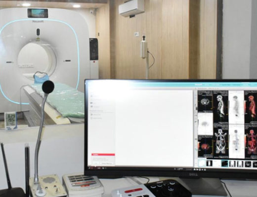
A PET (positron emission tomography) scan is a type of medical imaging that uses a radionuclide tracer to produce images of the body’s organs and tissues. Like a gamma camera, a PET scan involves the injection of a radionuclide tracer into the patient’s body. However, unlike a gamma camera, which detects gamma rays emitted by the tracer, a PET scan uses a detector to detect the annihilation of positrons, which are produced when the tracer decays.
The PET scan consists of a detector and a computer system that processes the data collected by the detector. The detector is typically an array of scintillation crystals, which are similar to the scintillator used in a gamma camera. The crystals are surrounded by photomultiplier tubes (PMTs), which are sensitive detectors that convert the light emitted by the crystals into an electrical signal. The electrical signal is then processed by the computer system to produce an image of the distribution of the tracer in the body.
PET scans are often used to detect cancer, evaluate the effectiveness of cancer treatment, and to diagnose neurological conditions. They are also used to assess the function of the heart and to evaluate the blood flow to the brain. PET scans are able to provide more detailed images of the body’s metabolism and biochemical processes than other imaging modalities, such as CT (computed tomography) or MRI (magnetic resonance imaging).
In summary, a gamma camera and a PET scan are both types of medical imaging that use a radionuclide tracer to produce images of the body’s organs and tissues. The main difference between the two is the type of detector used to detect the emitted radiation and the level of detail in the resulting images.
Also See: