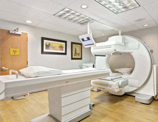
What is Gamma Camera?
Nuclear medicine requires the use of a gamma camera, sometimes referred to as a scintillation camera, as a diagnostic and monitoring tool. Radiopharmaceuticals, or radioactive drugs, are used in the medical speciality of nuclear medicine to diagnose and treat patients. Patients are given a little dose of this drug, which causes the body to emit a tiny amount of radiation for a brief period of time that may be detected by a gamma camera. The radiation exposure is negligible and has no negative effects on the person. Ionizing radiation released from the body is utilised by the gamma camera to produce pictures.
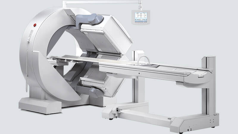
The camera heads employed by gamma cameras to record the pictures typically range from one to three. Each camera head has a flat surface that has to be placed as near to the patient as feasible. A gantry or strong metal arms are frequently used as camera head supports. The process is painless, and neither the camera nor the medicine dose given along with the radiation cause any discomfort. There has never been an issue with the process going wrong.
What is a Gamma Camera used for?
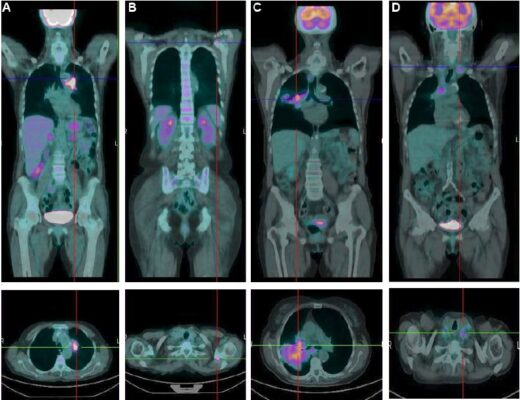
Gamma cameras are crucial for radiologists because they allow them to perform “Scintigraphy scans,” as they are known in medicine, which provide detailed information about the operation of a variety of organs, including the lungs, heart, thyroid, parathyroids, liver, kidneys, and adrenals, to name a few. Because they are used to diagnose medical disorders and track post-treatment progress, they are of utmost use in medical care. They can detect the hot areas caused by the spread of malignancy to bones or organs.
The scans that were collected in this way aid specialists in the diagnosis and identification of different malignancies, congenital anomalies, and other illnesses, enabling them to formulate a successful action plan. Specialists are able to assess how well patients respond to continuous therapies, which opens the door to corrective action to alter and adjust treatment programmes.
How does a Gamma Camera Work?
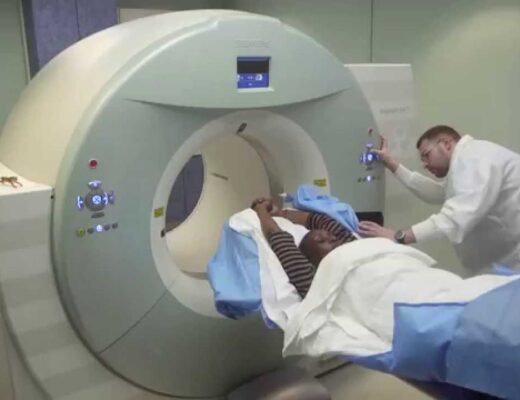
The patient is first given a tracer or radiopharmaceutical intravenously, orally, orally, or by inhalation. Gamma cameras may provide photos when radioactive tracers are present in the patient’s body for a limited time. Technetium-99m is the most widely used tracer, and these cameras capture radiation coming from it. It may be integrated into a number of molecules and has a reasonably long half-life of six hours, allowing it to target many bodily systems. A crystal in the camera that sparks or scintillates (flashes) in reaction to exposure to gamma rays may detect and track radiation that the tracer emits as it moves through the human body.
The crystal in the camera is placed in front of a collection of light sensors so that, upon exposure, the flashes of light transform into an electrical signal. Because of this, the device is sometimes referred to as a scintillating camera. Because specialists can identify diseases based on the accumulation or exclusion of tracer in particular regions, this technique aids in gauging the functioning and health of numerous human systems and organs.
The gamma camera is distinct from other imaging technology because it evaluates numerous body functions in addition to anatomy and structure.
Prior to the information age, gamma cameras may not have been particularly useful, but today’s computer technology allows for the quick computation of complex computations, turning the radiation detected by the camera into useful information for radiologists. In reality, because the pictures are produced in a fraction of a second, the spread of the radioisotope through the patient’s body may be monitored in real time. This implies that medical professionals may observe extremely detailed pictures demonstrating a process, such as the heart contracting, rather than merely static views of an organ or structure. Dynamic Imaging is the name given to this kind of imaging.
Prior to the information age, gamma cameras may not have been particularly useful, but today’s computer technology allows for the quick computation of complex computations, turning the radiation detected by the camera into useful information for radiologists. In reality, because the pictures are produced in a fraction of a second, the spread of the radioisotope through the patient’s body may be monitored in real time. This implies that medical professionals may observe extremely detailed pictures demonstrating a process, such as the heart contracting, rather than merely static views of an organ or structure. Dynamic Imaging is the name given to this kind of imaging.
Who Operates a Gamma Camera?
Several medical and technical experts with training in radiology or nuclear medicine operate and manage gamma cameras. In addition to professionals that specialise mainly in Radiology, many specialists have expertise in both nuclear medicine and cardiology or cancer. Nuclear medicine technologists actually operate the equipment, but other professionals engaged in the patient’s treatment and diagnosis will decide whether gamma camera testing and monitoring is necessary. Healthcare professionals with a degree in nuclear medicine are known as nuclear medicine technicians.
They are now qualified to administer injections, take blood samples, and utilise and quantify radiopharmaceuticals. This indicates that they are the experts who additionally inject the tracer before the gamma camera is operational. They have the knowledge and abilities to process and interpret the results of tests and nuclear medicine research in addition to being permitted to utilise gamma cameras. Additionally, they are aware of the many illnesses and medical issues that nuclear medicine treats.
What is the duration to complete a Nuclear Scan with Gamma Camera?
Scintigraphy with Technetium 99 usually takes 3-4 hours. There are two parts to it. In the first part a radio-isotope tracer is injected in the vein and in the second part the uptake is captured by the camera.
The total duration of the scan depends on the Radiopharmaceutical used. A Bone scan may take 1 to 4 hours.
How is Safety Ensured while using a Gamma Camera?
Patients are exposed to some amount of radiation during such procedures, but the amount varies depending on the specific procedure, the organ or structure being examined and the physical dimensions of the patient. Either way, health care providers follow stringent safety practices and will only administer the lowest possible dosage that can provide high quality results, so as to minimize unnecessary exposure to the drug.
Also See: